
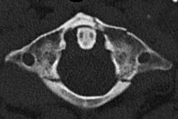
The ring-shaped Atlas (C1) supports the head. Together with the Axis (C2), these cervical vertebrae form the cranio-cervical junction. The occipito-atlantal and atlanto-axial articulations account for about 50% of the flexion and rotation in the cervical spine. Due to their high degree of mobility, these bony segments frequently show fractures in adults after spinal trauma. Although Atlas fractures rarely require surgery, they should be promptly recognized and treated in all patients. Every bony fracture of the Atlas warrants a thorough examination of the ligamentous structures between Occiput-C1 and C1-C2. Up to 70% of Atlas fractures may involve other spinal elements, especially C1-C2 complex fractures.
Epidemiology
Atlas fractures account for 2-3% of all spinal injuries and about 7-13% of all cervical vertebra fractures (~25% of all injuries at the cranio-cervical junction). The average age of patients with Atlas fracture is 64 years, with a bimodal distribution (20-30 years and 80-85 years). The most common type is an isolated anterior or posterior arch fracture (Gehweiler Type I & II). The second most common are isolated burst fractures (Gehweiler Type III & IV) without neurological deficits.
Clinical presentation
C1 fractures typically present with neck pain or torticollis without signs of neurological dysfunction. In unstable fracture types, spinal cord compression may clinically manifest as neurological deficits, especially of the lower cranial nerves (VI to XII).
Radiological findings & Classification

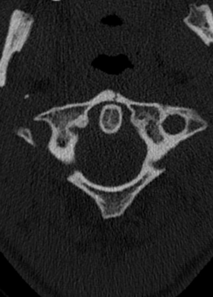

Several classification systems exist for Atlas fractures. The three most commonly used in clinical studies are the Jefferson, Landells, and Gehweiler classifications. The Jefferson classification is mainly used in the US and Asia, while the Gehweiler classification is preferred in Europe.
Gehweiler Classification
- Type I: Fracture of the anterior arch (uni- or bilateral)
- Type II: Fracture of the posterior arch (uni- or bilateral)
- Type III: Combined fractures of anterior & posterior arch (= Jefferson Fx = “burst” Fx)
- Type IV: Fracture of the lateral mass of the Atlas
- Type V: Fracture of the transverse process of the Atlas.
Transverse Atlantal Ligament (TAL) Injuries – Dickman Classification
- Type 1a: Central TAL rupture
- Type 1b: Rupture near the lateral mass
- Type 2a: Bony avulsion at the ligamentous attachment
- Type 2b: Bony avulsion with fracture of the lateral mass.
Jefferson Fracture
Combined fractures of the anterior and posterior Atlas arch are also called Jefferson fractures (= Gehweiler Type III). Professor Geoffrey Jefferson first described these combination fractures of the Atlas in 1920 and further subdivided them. The classic Jefferson fracture is the 4-part fracture, where both the anterior and posterior arches of the Atlas are broken on both sides. Fracture types with 2 or 3 parts can also occur. Jefferson fractures result from axial forces on the cervical spine (e.g., diving into shallow water). The forces are transmitted directly from the occipital condyles through the joint surfaces to C1 and C2. Due to the absence of intervertebral discs, there is no cushioning effect, and the forces are laterally dissipated due to joint positioning, leading to bursting of the Atlas arches and tearing of the transverse atlantal ligament (TAL). This can result in C1-C2 instability with spinal cord compression.
Rule of Spence
In the “AP-Dens view with open mouth,” asymmetries can be detected if there is displacement of Atlas fracture fragments. According to the “Rule of Spence,” rupture of the transverse atlantal ligament is highly likely if the combined lateral overhang of both lateral masses of C1 on C2 exceeds 6.9 mm. Heller et al. set the cutoff at >8.1 mm. However, MRI of the cervical spine provides better assessment of ligamentous structures and functional imaging.


Indication for surgery
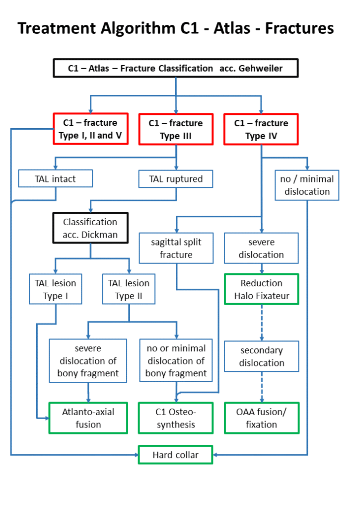
Currently, there are no universally valid treatment guidelines based on Level-I studies. Treatment ranges from non-operative to operative management, depending on fracture pattern and integrity of surrounding ligaments. These fractures pose a unique dilemma in elderly patients due to lower healing capacity and comorbidities. Additionally, combination or complex fractures (C1-C2), which can occur in up to 70% of Atlas fractures, limit general indications for conservative or operative management. The German Society for Orthopaedics and Trauma (DGOU) has developed guidelines (as of 2018) for the treatment of isolated C1 fractures, based only on medical evidence levels III-IV due to limited data. Similar guidelines have been developed by AO Spine. A summary comparison is shown in flowcharts in the article.
Outcome
Most studies on non-operative management of C1 – Atlas – fractures have been conducted in patients with Jefferson fractures (= Gehweiler Type III). The overall prognosis is good for stable fractures, and in most cases, conservative treatment leads to adequate fracture healing. In patients with occult fractures, poor healing, or bony anomalies, the “non-fusion” rate is about 17% to 20%.
References
- ???