
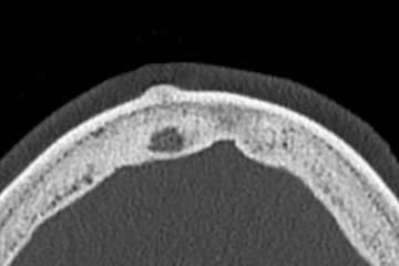
Osteoma is the most common primary bone tumor of the calvaria. The tumor grows slowly and is benign. It can also occur in the mastoid, the paranasal sinuses, and the mandible.
Epidemiology
Osteomas occur more frequently in women. The peak age is between 50 and 60 years. As part of Gardner’s syndrome, it is part of the triad: I: Multiple cranial osteomas, II: Polyposis of the colon (adenomas), and III: Connective tissue tumors (atheromas, lipomas, fibromas, leiomyomas).
Clinical presentation
Osteomas are usually asymptomatic. When osteomas occur in the paranasal sinuses, chronic or recurrent sinusitis may develop. When located in the forehead area or are large in size, they may present a cosmetic problem, especially in women.
Radiological findings
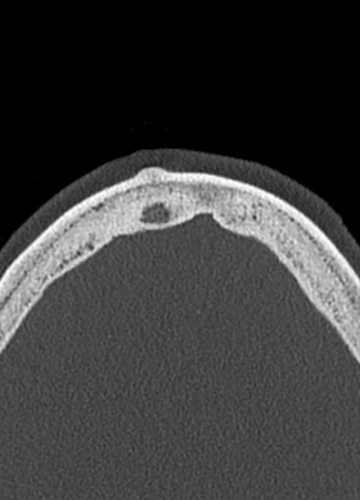
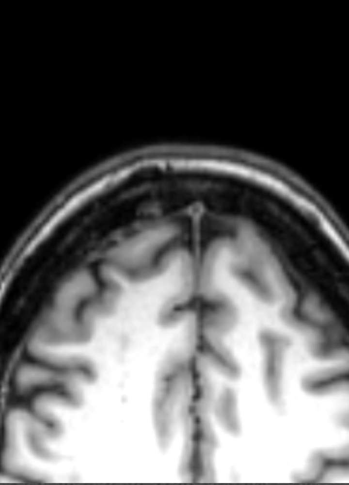
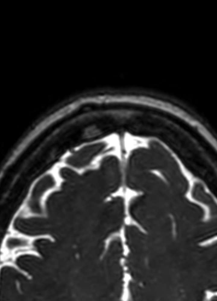
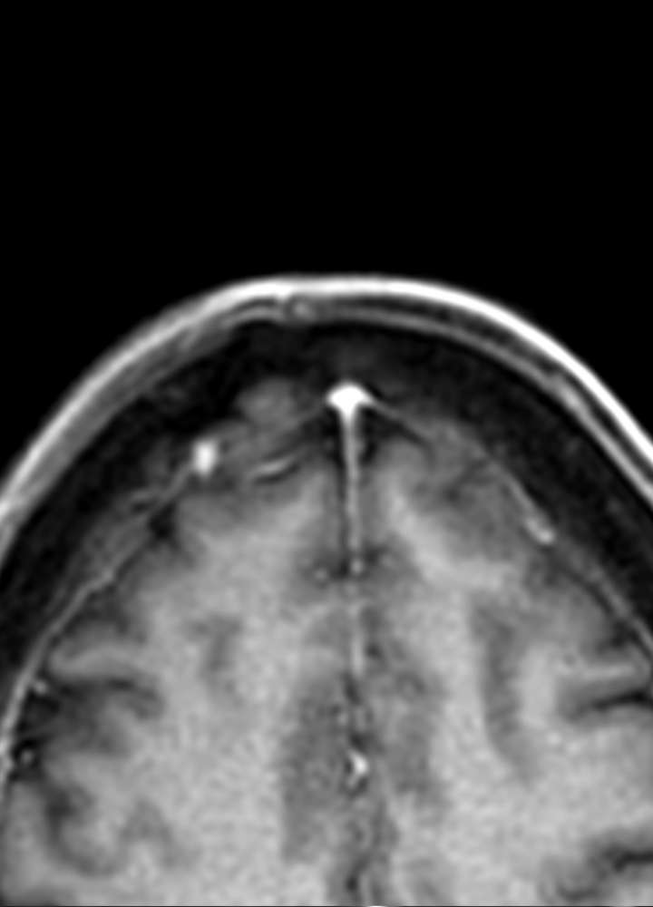
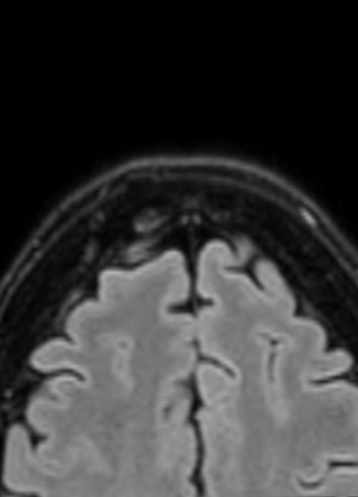
Osteomas are typically round, sclerotic, and well-delineated from the surrounding bone. The origin is usually the external cortical bone. Occasionally, osteomas may appear spongiform and somewhat softer. The venous structures remain intact (differential diagnosis to meningioma). Osteomas do not take up contrast medium on MR imaging. In PET-CT / MR imaging, they appear as “hot” nodules.
Differential diagnoses
- Metastases (osseous, osteoblastic)
- Fibrous dysplasia
- Calvarial impression fracture
- Paget’s disease
- Osteoblastoma
- Osteosarcoma
- Tuberous sclerosis
Indication for surgery
Symptomatic patients: If there are symptoms due to the osteoma (e.g., pain, pressure, dysfunction), complete surgical removal (“total resection”) is indicated to relieve symptoms and prevent recurrence.
Cosmetic reasons: If the osteoma, particularly in the forehead area, poses an aesthetic issue (common in women), surgery can also be performed. The entire osteoma can be removed, or if possible, just the external cortical bone can be removed to improve cosmetic results.
Asymptomatic osteomas: If there are no symptoms or cosmetic issues, immediate surgery is not necessary. In such cases, regular monitoring (e.g., through imaging) is recommended to detect any changes early.
The choice of procedure depends on the size, location, and extent of the osteoma, as well as the individual needs of the patient. Resection can be performed via craniotomy, followed by plastic covering with bone cement. Alternatively, only the outer bone layer (external cortical bone) can be removed (shaved) to improve the cosmetic outcome.
Outcome
Very good, recurrences are rare with complete excision.