
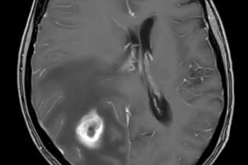
Cerebral metastases account for up to 50% of all brain tumors in adults. Approximately 15–30% of patients with malignant disease develop brain metastases during the course of progressive systemic disease. About 90% of patients with a first diagnosis of brain metastasis already have additional extracranial metastatic disease at the time of diagnosis. In patients without a known history of malignancy, neurological symptoms caused by brain metastases represent the initial manifestation of cancer in approximately 15% of cases. Metastatic spread to the brain occurs predominantly via the hematogenous route.
Epidemiology
Brain metastases are the most common intracranial tumors in adults, accounting for slightly more than 50% of all intracranial pathologies. Among supratentorial metastases, approximately 80% are located in the cerebral hemispheres, typically at the gray–white matter junction, most frequently in the postcentral region at the border of the parietal, temporal, and frontal lobes. This distribution is thought to reflect the terminal vascular territory of the middle cerebral artery.
In the posterior cranial fossa, metastases are the most common tumors in adults, likely due to hematogenous spread via the Batson venous plexus, a valveless venous network allowing retrograde tumor dissemination.
Lung and breast carcinomas together account for more than 50% of all brain metastases. In children, metastases represent only about 6% of brain tumors.
Etiology of cerebral metastases in adults
| Primary Tumor | Proportion |
|---|---|
| Lung carcinoma | 44% |
| Breast carcinoma | 10% |
| Renal cell carcinoma | 7% |
| Gastrointestinal tract tumors | 6% |
| Melanoma | 3–16% |
| Other tumors | 10% |
Clinical presentation
As with most brain tumors, neurological deficits typically develop subacutely over weeks to months, rather than acutely as seen in vascular events. Common symptoms include:
- Headache (approximately 50% of cases), often accompanied by nausea and vomiting
- Focal neurological deficits (e.g., hemiparesis, aphasia, visual disturbances)
- Epileptic seizures (approximately 15%)
- Neuropsychiatric and personality changes, including depression, lethargy, apathy, and confusion
Caution: Acute neurological deterioration may occur in the setting of hemorrhagic metastases.
Radiological findings


On MRI, brain metastases exhibit a wide range of appearances. They may be solid or cystic and show homogeneous or heterogeneous signal characteristics. Calcifications are rare. Lung, breast, and gastrointestinal carcinomas tend to form cystic metastases.
Metastases typically demonstrate contrast enhancement, which may be nodular or ring-like, often surrounding a centrally necrotic or hemorrhagic core. Certain tumor types – including melanoma, renal cell carcinoma, breast carcinoma, lung carcinoma, thyroid carcinoma, retinoblastoma, and choriocarcinoma – have a strong tendency toward intratumoral hemorrhage.
Prostate carcinoma and hormone receptor–positive breast carcinoma frequently metastasize to bone before involving the brain parenchyma. All cerebral metastases are associated with peritumoral vasogenic edema, often disproportionate to lesion size.
Compared with astrocytomas, metastases are usually better demarcated from the surrounding brain parenchyma.
Caution: In cases of atypical intracerebral hemorrhage, vascular malformations and hemorrhagic metastases must be considered in the differential diagnosis.
Differential diagnoses
- Primary or secondary lymphoma
- Gliomas
- Meningioma (particularly for dural-based metastases)
- Hemorrhagic occult cerebrovascular malformations (e.g., cavernoma)
- Brain abscess
Special considerations
Solitary metastases
At initial presentation, approximately 50% of patients appear to have a solitary brain metastasis on CT imaging. With additional MRI, the proportion of truly solitary lesions decreases to less than 30%. Solitary metastases are found in the brainstem in approximately 1–3% of cases.
Increasing incidence of brain metastases
The incidence of brain metastases is rising, likely due to prolonged survival of cancer patients resulting from improved systemic therapies. Advances in neuroimaging have also increased detection rates. Additionally, systemic chemotherapy may transiently weaken the blood–brain barrier, facilitating cerebral dissemination. However, many chemotherapeutic agents still do not adequately penetrate the blood–brain barrier.
Small Cell Lung Cancer (SCLC)
SCLC accounts for approximately 20% of lung cancers, while 80% are non-small cell lung cancers (NSCLC). SCLC exhibits a strong propensity for brain metastases: up to 80% of patients who survive longer than two years develop cerebral metastases. Treatment typically includes radiotherapy and chemotherapy, as SCLC is highly radiosensitive. Surgical resection is reserved for life-threatening large lesions or local recurrences after radiation therapy.
Non-Small Cell Lung Cancer (NSCLC)
NSCLC generally carries a better prognosis than SCLC. Due to its moderate resistance to radiotherapy, surgical resection is often the preferred treatment for accessible lesions.
Melanoma
Symptomatic brain metastases from melanoma typically appear approximately 14 months after diagnosis of the primary tumor. Median survival after diagnosis of cerebral metastases is around 6 months. Surgical resection of 1–4 metastases is indicated if complete resection is feasible and systemic disease progression is slow. Whole-brain radiotherapy (WBRT) provides palliative benefit, extending survival by approximately 2–3 months. Stereotactic radiosurgery (SRS) is used for lesions <3 cm when surgery is not feasible and when no more than four lesions are present. For multiple non-life-threatening metastases, systemic therapy—including alkylating agents, immunomodulators, BRAF inhibitors, and monoclonal antibodies—is preferred.
| Radiosensitivity | Tumor Types |
| Highly sensitive | SCLC, lymphoma, leukemia, multiple myeloma, germ cell tumors |
| Moderately sensitive | Breast cancer |
| Moderately resistant | Colon cancer, NSCLC |
| Highly resistant | Thyroid cancer, renal cell carcinoma, malignant melanoma, sarcoma, adenocarcinomas |
Prognostic factors associated with improved outcome
- Karnofsky Performance Score (KPS) > 70
- Age < 60 years
- Brain metastases without extracranial disease
- Systemically controlled primary tumor
- Survival > 1 year since initial cancer diagnosis
- Solitary or limited number of brain metastases
- Female sex
Indication for surgery
In patients with an unknown primary tumor, stereotactic biopsy is recommended for diagnostic confirmation unless complete surgical resection is feasible. In cases of uncontrolled systemic disease, poor clinical status (KPS <70), and limited life expectancy, biopsy followed by palliative WBRT – or best supportive care – may be appropriate.
In patients with systemic disease control and KPS >70, surgical resection should be pursued if lesions are completely resectable and limited in number (<4). In pancerebral metastases (>4 lesions), surgery is reserved for life-threatening mass effect or severe neurological symptoms. Adjuvant radio-chemotherapy is mandatory.
For solitary lesions:
- Controlled systemic disease
- Complete surgical resectability
- Symptomatic or life-threatening lesion
- Radiation-resistant tumor
- Recurrent SCLC lesion
- Unknown primary tumor (biopsy or resection)
For multiple lesions:
- One lesion is symptomatic or life-threatening
- All lesions (<4) are completely resectable
- No prior histological confirmation (biopsy or resection)
Outcome
With optimal treatment and a Karnofsky score >70, median survival across all brain metastasis groups is approximately 26–32 weeks (6–8 months). Without treatment, median survival after onset of neurological deficits is about 1 month. Steroid therapy alone extends survival to approximately 2 months. WBRT combined with steroids results in a median survival of 3–6 months.
Patients with a solitary brain metastasis and no evidence of active systemic progression demonstrate the best outcomes. Complete surgical resection followed by WBRT yields a median survival of approximately 40 weeks (10 months), although 22% experience recurrence within one year. Treatment with SRS plus WBRT offers comparable local tumor control but less predictable tumor size reduction. Patients with fewer than four completely resected metastases show survival rates similar to those with resection of a single metastasis.
References
- ???